


Skin Cancer Types
Malignant melanoma is the deadliest form of skin cancer. When detected early and treated appropriately by a dermatologist the survival rate is 98%. The cause is unclear but is related to overexposure to the UV radiation from the sun and tanning beds. However, it can also develop on skin that has only been minimally exposed to UV radiation.
Nonmelanoma skin cancers include basal cell and squamous cell carcinomas. They are the most common types of skin cancer. They are also caused by overexposure to UV radiation from the sun and tanning beds. However, squamous cell carcinoma can develop on areas of the body that have not been exposed to the sun; and it can spread to the lymph nodes. Basal cell carcinoma does not spread to other areas of the body. More than a 95% of nonmelanoma skin cancers can be cured. However, they can become disfiguring if not caught early.
Basal cell carcinoma looks like a small, raised, pearly, pink or white scaly patch that may bleed; or an open sore that doesn’t heal. Squamous cell carcinoma looks like thick, rough, scaly patches that may crust or bleed.
What are the ABCDEs?
The ABCDEs of skin cancer are key warning signs of potentially malignant lesions and changes that can help detect melanoma and nonmelanoma skin cancers early while they can be cured. It is important to note that not all melanomas originate in moles.
Examine your skin regularly. Look at sun exposed skin as well as skin that is protected by clothing. Observe any new skin lesions and changes to existing skin lesions using the ABCDEs. In addition, examine any growth that itches, bleeds, or won’t heal. If you find any spot that shows the ABCDEs or bleeds and won’t heal, contact Dr. Sheila Farhang for a professional opinion.
- A Asymmetrical Shape. Normal moles and colored growths are usually symmetrical (even on both halves like a circle) and are smaller than ¼ inch in size. A hallmark of melanoma is a mole that changes from a symmetrical shape to an irregular shape. A change in shape is just one feature of a potentially malignant growth.
- B Borders. Blemishes and marks are typically round or oval and have smooth borders. Ill-defined or blurry borders can be a sign of a precancerous growth or cancer.
- C Colors. Benign moles are usually uniform in color. A mole with more than a single color is suspicious. Melanomas usually show a mix of two or more colors or shades of brown and black, or it may be red or blue.
- D Diameter. Melanomas are larger than most moles. A mole or growth that is larger than a quarter of an inch in diameter suggests melanoma.
- E Evolution. Benign moles look the same over time. A mole that is evolving its shape, color, or size or that develops a new symptom like bleeding, itching or crusting is more likely to be dangerous.
Skin Cancer Screening
Dr. Farhang strongly recommends annual skin cancer screenings to ensure early detection. Early detection, before symptoms appear, makes skin cancer easier to treat and provides the best outcome. If screening reveals a problem, diagnostic tests will be needed.
Consultation
During your consultation, Dr. Farhang will ask about your symptoms including any changes in size and appearance of a mole or other skin lesion, and whether the growth is itchy, painful or bleeds. She will ask if there is a family history of skin cancer, your risk factors such as how many sunburns you have had, and whether you have had skin cancer before.
She will conduct a physical examination of the growth taking into consideration its appearance and your symptoms. She will examine the rest of your body to assure all potential tumors are identified. She may also feel the lymph nodes in the neck, underarm and groin near the tumor if deemed necessary.
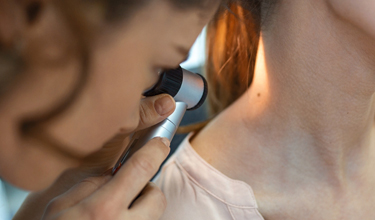
During the physical exam, Dr. Farhang will use a special microscope called a dermatoscope to examine and evaluate all questionable growths. Dermoscopy improves the accuracy of a diagnosis and assesses the need for a biopsy.
Skin Biopsy
A skin biopsy is the best and often the only test necessary to diagnose skin cancer. All biopsies are performed under local anesthetic for your comfort. During the biopsy a part of the growth or the entire growth will be removed. The biopsied tissue will be sent to a laboratory for diagnosis. You may have a sore or stitches after the biopsy. Dr. Farhang will provide wound care instructions and the office will notify you of the results of the biopsy.
When a skin cancer is diagnosed, Dr. Farhang will order additional diagnostic tests such as blood tests and imaging tests like a CT scan, x-ray and/or MRI. Because melanoma and squamous cell carcinoma can spread to the nearby lymph glands, Dr. Farhang will feel your lymph glands and if they are enlarged or swollen, she may order a lymph node biopsy. Enlarged lymph glands may suggest further diagnostic testing to rule out metastasis.
Dr. Farhang is a board – certified dermatologist and double-fellowship trained Mohs micrographic surgeon, cutaneous surgeon, and reconstruction and cosmetic surgeon. When she has made the diagnosis of melanoma, the next steps are staging and grading the tumor.
Grading and staging the tumor
Using the information from your exam, physical and biopsy, Dr. Farhang will grade the tumor to determine how abnormal the cells and tissues look under a microscope. This indicates how quickly the tumor may grow and spread. The more abnormal the cells, the more likely the cancer will grow and spread. Next the tumor is staged to determine how large the tumor is and how far it has spread.
Grading and staging are important for treatment planning and predicting outcome or prognosis.
Schedule a skin cancer screening consultation
Dr. Sheila Farhang is a board-certified medical dermatologist and Mohs surgeon; and the founder and medical director of Avant Dermatology and Aesthetics in Tucson, Arizona. She has extensive training in cutaneous oncology, and trained at Moffitt Cancer center, one of the top cancer centers in the country. She couples her exceptional knowledge, skills and experience, with state-of-the-art technology to provide the best dermatology care available. If you’re concerned that you may have skin cancer, do not wait, schedule a skin cancer screening today.

 / 291 Reviews
socialize with avant
#naturalskin